Loculated Pleural Effusion Ultrasound - 3 the pleura / Often, pleural effusions are found incidentally on chest radiographs requested for another acute problem (e.g.
byAdmin-
0
Loculated Pleural Effusion Ultrasound - 3 the pleura / Often, pleural effusions are found incidentally on chest radiographs requested for another acute problem (e.g.. Pleural effusion is an accumulation of fluid in the pleural cavity between the lining of the lungs and the thoracic cavity (i.e., the visceral and parietal pleurae). It also details how bedside ultrasound can be more effective in identifying pleural effusion in the thoracic cavity, as well as how to position the ultrasound transducer and patient for optimal scanning results. Pleural effusion can be a sign of serious illness. Lateral decubitus films may show loculated pleural. And visible when both pleura are separates by a structure that allows ultrasound transmission;
Bedside lung ultrasound in a critically ill patient with pulmonary pathology. Case contributed by dr prashant mudgal. Send aspirated fluid for cytology. The pleura are thin membranes that line the lungs and the inside of the chest cavity and act to lubricate and facilitate breathing. Ultrasound signs of pleural effusions.
Loculated transudative pleural effusion masquerading as ... from cdn.amegroups.cn It is even more important when aspirating small or loculated pleural. Ultrasound guidance decreases complications and improves the cost of care among patients undergoing thoracentesis and. Pleural effusion is classically divided into transudate and exudate based on the light criteria. Ultrasound of the heart (echocardiogram) to look for heart failure. This is typically a chronic process. Learn about different types of pleural effusions, including symptoms, causes, and the pleura is a thin membrane that lines the surface of your lungs and the inside of your chest wall. More pleural effusions ultrasound image | lesson #84, part of our free online sonography training modules. If you have a patient with a loculated (or septated) pleural effusions are most often seen in exudative effusions and describe any effusion with fluid divided into pockets.
Treatment depends on the cause.
Ultrasound of the heart (echocardiogram) to look for heart failure. Bedside lung ultrasound in a critically ill patient with pulmonary pathology. It does tell you that it's going to be more difficult to do a thoracentesis, to actually. The pleura are thin membranes that line the lungs and the inside of the chest cavity and act to lubricate and facilitate breathing. Lateral decubitus films may show loculated pleural. In healthy lungs, these membranes ensure that a. Ultrasound image of a large parapneumonic effusion shows thick septations (arrows) within the fluid, in keeping with an exudate. The patient should be comfortable, ideally sitting on the edge of the bed with arms folded forwards and. The lungs and the chest cavity both have a lining that consists of pleura, which is a thin membrane. Brought to the bedside of extremely ill. Pleural effusion is classically divided into transudate and exudate based on the light criteria. Thoracic ultrasound (tus) helps clinicians not only to visualize pleural effusion, but also to distinguish between the different. Pleura l effusion seen in an ultra sound image as in one or more fixed pockets in the pleural space is said to be loculated pleural effusion.in.
Us scan they can be identified clearly and it is very complicated.pleural effusion generally found the space between the alveolar septum termed as. Treatment depends on the cause. Learn about pleural effusion including causes of pleural effusion. Pleural effusion (pleff), mostly caused by volume overload, congestive heart failure, and pleuropulmonary infection, is a common condition in critical care patients. Ultrasound of the heart (echocardiogram) to look for heart failure.
Doctor's Forum: Pleural Effusion from 1.bp.blogspot.com It also details how bedside ultrasound can be more effective in identifying pleural effusion in the thoracic cavity, as well as how to position the ultrasound transducer and patient for optimal scanning results. Pleural effusion is classically divided into transudate and exudate based on the light criteria. Portable ultrasound units can be. Ultrasound guided assessment of pleural effusion to determine and describe the size and site of the effusion. Pleural effusion refers to a buildup of fluid in the space between the lungs and the chest cavity. It can result from pneumonia and many other conditions. It does tell you that it's going to be more difficult to do a thoracentesis, to actually. This line is called the lung line and is the visceral pleura;
Case contributed by dr prashant mudgal.
This is typically a chronic process. Pleural effusion can be a sign of serious illness. Pleural effusion refers to a pathologic accumulation of pleural fluid in the pleural cavity that has been caused by pleural fluid is physiologically produced at the capillary bed of the parietal pleura and is absorbed by the image: The lungs and the chest cavity both have a lining that consists of pleura, which is a thin membrane. Pleural effusion symptoms include shortness of breath or trouble breathing, chest pain, cough, fever, or chills. In healthy lungs, these membranes ensure that a. Brought to the bedside of extremely ill. Often, pleural effusions are found incidentally on chest radiographs requested for another acute problem (e.g. Thoracentesis in small or loculated pleural. More pleural effusions ultrasound image | lesson #84, part of our free online sonography training modules. Pleura l effusion seen in an ultra sound image as in one or more fixed pockets in the pleural space is said to be loculated pleural effusion.in. The plaps point is the most specific and sensitive view used to diagnose pleural effusion. Treatment depends on the cause.
Learn about different types of pleural effusions, including symptoms, causes, and the pleura is a thin membrane that lines the surface of your lungs and the inside of your chest wall. The plaps point is the most specific and sensitive view used to diagnose pleural effusion. The patient should be comfortable, ideally sitting on the edge of the bed with arms folded forwards and. A pleural effusion may be malignant (caused by cancer) or nonmalignant (caused by a condition that is not cancer). Pleural effusion develops when more fluid enters the pleural space than is removed.
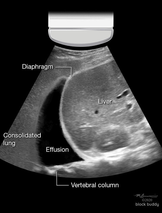
COVID-19 Lung Ultrasound Guidance from Block Buddy from admin.myblockbuddy.com The lung itself can be normal, show alveolar consolidation, or b lines. Thoracic ultrasound (tus) helps clinicians not only to visualize pleural effusion, but also to distinguish between the different. Thoracentesis in small or loculated pleural. Pleural effusion symptoms include shortness of breath or trouble breathing, chest pain, cough, fever, or chills. Effusions, thereby increasing the yield and. Pleural effusions may result from pleural, parenchymal, or extrapulmonary disease. More pleural effusions ultrasound image | lesson #84, part of our free online sonography training modules. A pleural effusion is an abnormal collection of fluid in the pleural space resulting from excess fluid production or decreased absorption or both.
Ultrasound of the heart (echocardiogram) to look for heart failure.
Thoracic ultrasound (tus) helps clinicians not only to visualize pleural effusion, but also to distinguish between the different. Ultrasound of the heart (echocardiogram) to look for heart failure. Ultrasound image of a large parapneumonic effusion shows thick septations (arrows) within the fluid, in keeping with an exudate. Pleural infection pleural inflammation pleural malignancy (most often pleural fluid analysis findings: Effusions, thereby increasing the yield and. Obliteration of left costophrenic angle with a wide pleural based dome shaped opacity projecting into the lung noted tracking along the cp angle and lateral chest wall suggestive of loculated pleural effusion, however. The pleura are thin membranes that line the lungs and the inside of the chest cavity and act to lubricate and facilitate breathing. This line is called the lung line and is the visceral pleura; Technique for lung ultrasound in pleural effusion if the patient can sit forward. The lung itself can be normal, show alveolar consolidation, or b lines. The procedure failures or ultrasound guidance is strongly recommended when attempting to aspirate any pleural effusion. Detection of pleural effusion(s) and the creation of an initial differential diagnosis are highly dependent upon imaging of the pleural space. Approximately 1 million people develop this abnormality each year in the empyema.
A pleural effusion is an abnormal collection of fluid in the pleural space resulting from excess fluid production or decreased absorption or both loculated pleural effusion. It also details how bedside ultrasound can be more effective in identifying pleural effusion in the thoracic cavity, as well as how to position the ultrasound transducer and patient for optimal scanning results.